
5 Surprising Facts About the Human Eye
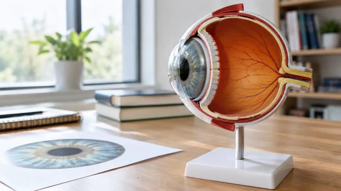
The human eye is routinely compared to an optical camera, yet its true biological reality is far more intricate and resilient. Rather than simply capturing static snapshots of the external world, the eye operates as a dynamic sensory extension of the central nervous system. It continuously captures variable light waves, refracts and filters those signals through living optical structures, translates photons into electrical impulses, and communicates instantaneously with the visual cortex to render our perception of reality.
Every waking second, an intricate matrix of specialized tissues, photoreceptor cells, fluid-filled chambers, and precision muscle groups collaborates to make human vision seamless. While everyday sight is often taken for granted, modern vision science reveals that the visual system possesses extraordinary capabilities—from distinguishing millions of subtle color gradients to rebuilding damaged tissue within hours.
Key takeaways
- The human eye can differentiate approximately 10 million distinct colors through the coordinated firing of three wavelength-sensitive cone photoreceptors.
- The brain reconstructs three-dimensional depth from flat surfaces using visual cues such as occlusion, texture gradient, and forced perspective.
- The cornea is among the fastest-healing tissues in the human body, capable of recovering from minor superficial abrasions in 24 to 48 hours.
- Human eyes are in perpetual motion, employing tiny involuntary movements called microsaccades to prevent static images from disappearing from view.
- Biometric patterns found in the iris and retina are entirely unique to each person, supporting iris recognition systems with error rates below 0.1%.
The Anatomical Architecture of Human Vision
To grasp the broader capabilities of sight, it is necessary to examine the primary biological hardware that makes optical processing possible. Vision is not generated by a single monolithic structure; rather, it relies on a synchronized relay of specialized tissues that direct and refine light as it passes through the eyeball.
The visual pathway begins at the cornea, the transparent, dome-shaped outer tissue at the front of the eye. Serving as the eye's foremost mechanical barrier, the cornea protects internal structures while performing a substantial portion of the eye's total optical focusing power. Immediately behind the cornea sit the iris and pupil. The iris, composed of pigmented connective tissue and delicate muscle fibers, contracts and dilates to adjust the aperture of the dark pupil at its center, regulating how much ambient light enters the ocular chamber.
Positioned directly behind the pupil is the crystalline lens. This clear, flexible structure alters its curvature to fine-tune the focus of light rays, projecting clean optical images onto the retina. The retina lines the interior back wall of the eyeball and contains millions of specialized photoreceptor cells known as rods and cones. Rods handle peripheral detection, motion, and vision in low-light environments, while cones operate in brighter conditions to resolve fine detail and color. The sensory impressions gathered by these photoreceptors converge at the optic nerve, a thick cable of nerve fibers that routes electrical signals straight to the visual processing centers of the brain.
Five Surprising Capabilities of the Human Eye
Vision researchers have spent centuries cataloging the physical limits and operational mechanisms of the eye. The following five attributes illustrate the remarkable precision, processing power, and biological durability of human sight.
Distinguishing Up to 10 Million Distinct Colors
Although human culture categorizes color across a familiar rainbow spectrum spanning red to violet, the human visual system can resolve approximately 10 million distinct colors. This exceptional range is made possible by three specialized types of cone cells embedded within the retina, each calibrated to respond to different wavelengths across the visible electromagnetic spectrum: short wavelengths (blue), medium wavelengths (green), and long wavelengths (red).

When light bounces off an object and enters the eye, these three cone populations fire simultaneous electrical impulses at varying intensities. The brain decodes the relative ratios of these incoming signals to construct a continuous spectrum of nuanced hues, tones, and saturations. This system is known as trichromatic vision. However, this perceptual range is not identical across the global population. Color vision deficiency alters how certain wavelengths are detected, creating difficulty when separating particular shades. This hereditary condition is documented in roughly 8% of men and 0.5% of women of Northern European descent. Outside the human realm, several animal species—including various birds, insects, and specific fish—possess four or more distinct cone types, enabling them to perceive ultraviolet spectrums and shades entirely invisible to people.
Perceiving Depth in Two-Dimensional Images
A remarkable computational feat of the visual system is its ability to extract three-dimensional depth from flat surfaces. Even when looking at a flat painting, photograph, or digital display, the brain applies specialized spatial rules to deduce the physical distances between depicted objects.
This process relies on monocular depth cues, including texture gradient, linear perspective, and occlusion. Occlusion happens when the profile of one object blocks or overlaps another; the brain immediately recognizes this overlapping boundary as definitive evidence that the partially obscured object sits farther away in physical space. These depth interpretations are indispensable for survival tasks such as driving, where operators must rapidly evaluate changing distances between vehicles and roadside obstacles. The reliance on these mental shortcuts is famously highlighted by optical illusions such as the Ames room illusion. By intentionally distorting the walls and floor while preserving a viewing peephole, the Ames room tricks the brain's perspective assumptions, causing two people of equal height to look like a miniature figure and a giant standing within the same space.
Rapid Cellular Healing and Tissue Regeneration
The human eye is one of the fastest-healing organs in the entire human body, displaying an extraordinary capacity to repair surface trauma. A minor superficial scratch or abrasion to the protective epithelial layer of the cornea can heal completely within 24 to 48 hours.
The human eye heals faster than almost any other organ in the body, with minor corneal abrasions routinely repairing themselves within 24 to 48 hours.
This rapid regenerative cycle is driven by the cornea's profound density of nerve endings and its immediate access to oxygen and metabolic nourishment, which fuel accelerated cellular division across the surface. While minor scrapes close quickly without intervention, deep lacerations, chemical injuries, or chronic infections can cause permanent scarring and vision impairment. For severe injuries, advanced ophthalmic procedures offer avenues for sight preservation. Surgeons perform corneal transplants to exchange clouded or scarred tissue with healthy donor tissue, while cutting-edge retinal implants are deployed to restore functional light perception for patients suffering from specific degenerative retinal diseases.
Continuous Motion Through Involuntary and Voluntary Scanning
Even when a person attempts to fix their gaze on a single pinpoint without blinking, the eye cannot remain motionless. The visual apparatus performs constant, microscopic adjustments known as microsaccades. These microscopic tremors shift the retinal field slightly, refreshing the incoming light on photoreceptors and preventing neural adaptation, a phenomenon where completely stationary visual stimuli fade entirely from conscious perception.
Beyond these micro-movements, the ocular system relies on several observable movement types to gather spatial data. Large, rapid voluntary adjustments called saccades reposition the eyes between visual targets, enabling us to read lines of text or scan an unfamiliar room. Conversely, when monitoring an object in flight, the visual system engages smooth pursuit movements, locking onto moving targets such as a soaring bird or a passing car to keep the focal target centered on the retina. The scientific discipline of tracking and recording these eye patterns, known as oculography, offers clinicians and researchers an objective window into mental workload, cognitive focus, and neurological health.

| Movement Type | Control Mechanism | Primary Visual Function | Everyday Example |
|---|---|---|---|
| Microsaccades | Involuntary | Prevents visual adaptation and image fading | Staring fixedly at a single stationary dot |
| Saccades | Voluntary | Scans environment and shifts focal points rapidly | Reading text across a printed page |
| Smooth Pursuit | Voluntary / Involuntary | Tracks continuous movement smoothly across the visual field | Watching a baseball travel through the air |
Unique Biometric Signatures Across Every Individual
Much like fingerprints, the physical architecture of the human eye is entirely unique to each individual. The complex iris contains a labyrinth of crypts, furrows, rings, and pigment clusters that develop during gestation and remain fundamentally stable throughout adult life. These singular characteristics make the iris one of the most reliable biometric markers in the world, with modern iris recognition systems demonstrating an error rate under 0.1%.
Retinal scanning provides another distinct layer of biological identification by mapping the pattern of blood vessels lining the posterior eye. Beyond individual uniqueness, the external geometry of the eye reflects ancestral and evolutionary adaptations. Physical variations in eye aperture, orbital depth, and eyelid shape differ across populations; for instance, individuals of African and Asian ancestry often display more rounded eye contours compared to the elongated orbital profiles frequently observed in populations of European heritage.
The Step-by-Step Mechanics of the Visual Pathway
Understanding how incoming environmental light translates into conscious thought reveals the delicate collaboration between ocular physics and neural computation. The conversion from raw photons to meaningful visual scenes follows an exact physiological sequence.
- Light waves reflect off environmental surfaces and strike the clear, convex surface of the cornea, which refracts the incoming light to initiate focusing.
- Light travels through the pupil opening, where the radial and sphincter muscles of the iris contract or dilate to establish the ideal level of illumination.
- The light rays enter the crystalline lens, which flexes and adjusts its curvature to converge the light precisely onto the inner lining at the back of the eyeball.
- Photons hit the retina, activating rods and cones that convert raw light energy into biochemical impulses through specialized phototransduction pathways.
- These generated impulses flow through intermediate retinal nerve layers and aggregate into the bundled fibers of the optic nerve.
- The optic nerve transports the dense visual data across the neural highway directly to the brain's visual cortex, which interprets color, depth, edges, and movement to assemble an interactive image.
Daily Protocols to Preserve Visual Health
Because the external tissues of the eye are exposed directly to dry air, airborne particles, and screen radiation, maintaining daily ocular hygiene and protection is essential for preserving high visual acuity throughout life.
- Undergo periodic comprehensive eye evaluations: Comprehensive clinical examinations ensure that internal ocular pressure, retinal vascular integrity, and visual sharpness are assessed routinely by an eye care specialist.
- Equip protective eyewear for dangerous activities: Wear safety glasses or face shields during yard maintenance, woodwork, mechanical tasks, or competitive athletics to block flying debris and chemical splashes.
- Implement the 20-20-20 visual rest routine: During computer or smartphone use, take a visual break every 20 minutes by focusing on a stationary target situated at least 20 feet away for 20 seconds to ease ciliary muscle tension.
- Preserve tear film moisture: Maintain consistent whole-body hydration and place room humidifiers in dry interior spaces to prevent the tear film from evaporating prematurely.
- Wear certified ultraviolet-blocking sunglasses: Select lenses that offer comprehensive UV filtering to shield internal structures like the lens and retina from long-term solar radiation damage.
- Clean hands thoroughly before touching the eye area: Wash your hands with soap before touching the face, applying cosmetics, or inserting contact lenses to minimize the transfer of harmful bacteria to the cornea.
Common Eye Care Mistakes That Threaten Vision
Many preventable eye problems develop from ordinary routines that place unnecessary mechanical or biological stress on sensitive ocular tissues. Identifying and eliminating these habits is critical for preventing avoidable vision damage.
- Dismissing ongoing eye irritation: Although the cornea can repair superficial abrasions in 24 to 48 hours, unyielding redness, sharp pain, or light sensitivity should never be neglected, as minor lesions can escalate into destructive bacterial ulcers.
- Overnight wear of standard contact lenses: Sleeping with standard lenses in place suffocates the cornea by blocking ambient oxygen, sharply escalating the incidence of severe bacterial and fungal keratitis.
- Overusing decongestant redness-relief eye drops: Frequent use of chemical drops formulated to constrict surface blood vessels triggers rebound vasodilation, resulting in chronic red eyes and worsening dryness when the medication wears off.
- Neglecting contact lens storage case replacement: Storage containers collect bacterial biofilms over time; reusing cleaning solution or using old cases introduces dangerous microbes directly onto contact lenses.
- Vigorously rubbing irritated eyes: Rubbing the eyelids when dust or grit is trapped inside presses abrasive particles into the delicate corneal tissue, resulting in painful surface lacerations.
- Reading and working in substandard lighting: Performing precision near-vision tasks in dim light forces the internal focusing system to overwork, producing rapid muscle fatigue, blurred vision, and persistent strain headaches.
Frequently asked questions
How many colors can the human eye detect, and how does it see them?
The human eye can detect approximately 10 million distinct colors. This wide spectrum is made possible by three varieties of cone photoreceptor cells in the retina, which respond selectively to short, medium, and long light wavelengths. The brain interprets the relative strength of signals received from all three cone types simultaneously to generate our full color perception.
How quickly can a scratched cornea repair itself?
The cornea possesses an extraordinary healing speed, with minor superficial abrasions typically regenerating within 24 to 48 hours. Its rapid recovery is supported by a dense network of nerve endings and a reliable supply of oxygen and metabolic nutrients that support fast cellular turnover.
Why do our eyes make tiny movements when looking at a stationary point?
These tiny involuntary shifts are known as microsaccades. They continuously alter the exact position of light hitting photoreceptors in the retina. Without these microscopic movements, the nervous system would adapt to static stimulation, causing an unchanging visual scene to fade entirely from view.
How does the brain perceive 3D depth from a flat 2D picture?
The brain decodes depth on flat surfaces by relying on monocular cues, including texture gradients, perspective lines, and occlusion. When one visual element partially blocks another, the brain automatically recognizes occlusion as proof that the obstructed object is further away in three-dimensional space.
Why is iris recognition considered more reliable than many other biometrics?
The human iris features a complex and permanent pattern of furrows, ridges, and crypts that is mathematically unique to every individual. Because these physical features remain exceptionally stable over a person's lifetime, iris scanning technologies boast an error rate of less than 0.1%.
The Bottom Line
The human eye is an extraordinary feat of biological engineering, uniting sophisticated optics, rapid cellular repair, and lightning-fast neural translation. From distinguishing millions of subtle color gradients to shielding itself through continuous microscopic motion, our visual system performs demanding tasks with effortless grace. Because optimal vision depends on delicate physical structures like the cornea, lens, and retina, proactive habits—including resting your eyes during screen work, shielding against physical trauma, and seeking prompt medical attention for persistent discomfort—remain the most reliable ways to preserve your eyesight for a lifetime.